Understanding Dental X-Ray Tube Maintenance: Why Preventive Care Extends Equipment Life
Dental x-ray equipment represents a significant capital investment for any practice, and the x-ray tube is its most critical — and most expensive — component. Understanding how these tubes work, recognizing early warning signs of failure, and implementing proper maintenance protocols can dramatically extend equipment life and prevent costly downtime.
How Dental X-Ray Tubes Work
At its core, an x-ray tube is a vacuum tube designed to convert electrical energy into x-ray radiation. Understanding the basic physics helps explain why maintenance matters so much.
The Cathode
The cathode assembly contains a tungsten filament — similar in concept to an old incandescent light bulb, but engineered for extreme precision. When heated by electrical current, the filament releases electrons through thermionic emission. A focusing cup surrounds the filament, shaping the electron stream into a narrow beam directed at the anode.
Most dental tubes use a dual-filament cathode: a small filament for fine-detail work (periapical images) and a larger filament for broader exposures (panoramic). The filament you select determines the focal spot size — the area on the anode where electrons strike, which directly affects image resolution.
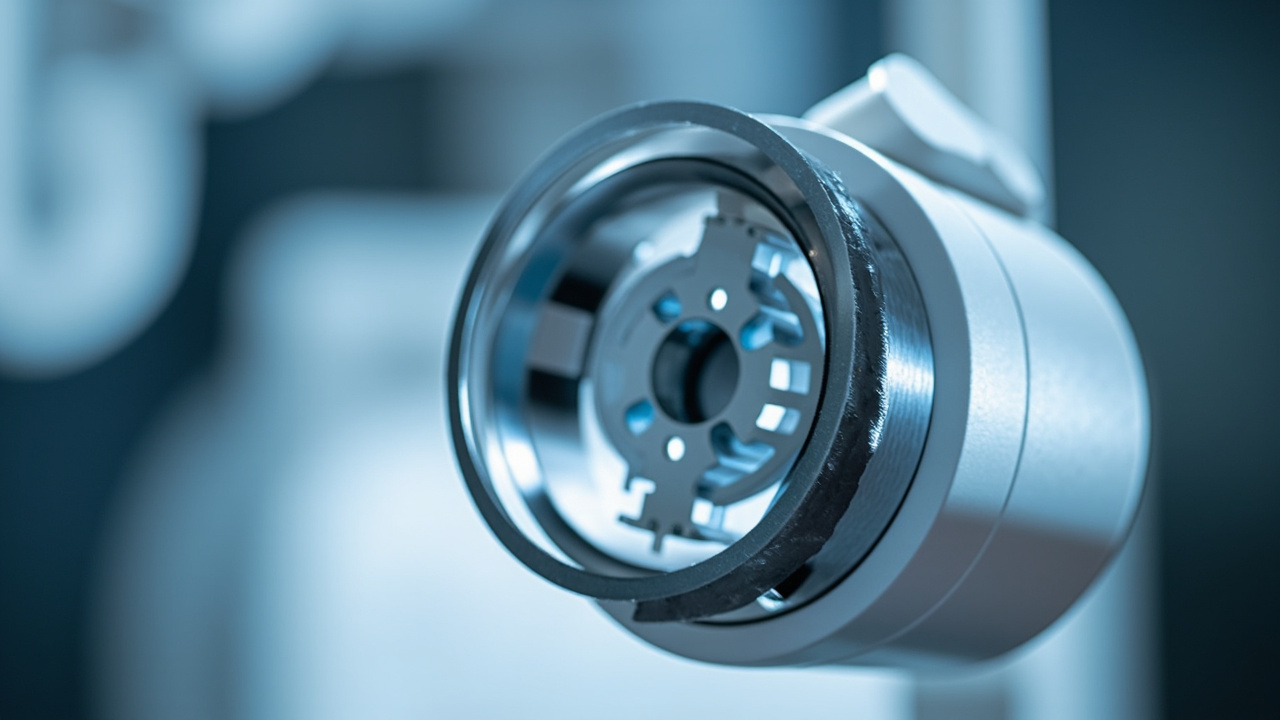
The Anode
The anode (or target) is where the magic happens — and where most of the punishment occurs. Electrons from the cathode slam into the anode’s tungsten target at tremendous speed. Only about 1% of that kinetic energy converts to x-rays; the remaining 99% becomes heat.
In dental intraoral units, you’ll typically find stationary anodes — a tungsten target embedded in a copper block that acts as a heat sink. Panoramic and CBCT units often use rotating anodes, where the tungsten disc spins at 3,000–10,000 RPM to distribute heat across a larger surface area.
The Focal Spot
The focal spot is the area on the anode where the electron beam strikes. A smaller focal spot produces sharper images but concentrates heat in a smaller area, accelerating wear. Dental intraoral tubes typically have focal spots of 0.4–0.7 mm, while panoramic units may use 0.5–1.0 mm spots.
The line-focus principle allows manufacturers to use an angled anode face to create an effective focal spot smaller than the actual area being bombarded — a clever engineering compromise between image quality and heat management.
Common Failure Modes
X-ray tubes don’t fail randomly. They wear out through predictable mechanisms, and understanding these helps you spot trouble early.
Anode Pitting and Roughening
Over thousands of exposures, the tungsten surface of the anode develops microscopic pits and roughness. This is the most common form of tube aging. As the surface degrades:
- X-ray output becomes less uniform
- Image contrast gradually decreases
- Heat dissipation becomes less efficient, accelerating further damage
- In severe cases, tungsten particles can vaporize and coat the glass envelope, causing electrical arcing
What accelerates it: Excessive exposure settings, inadequate warm-up, and exceeding duty cycle ratings. Making high-mA exposures on a cold tube is particularly destructive — thermal shock can crack the anode surface.
Bearing Wear (Rotating Anode Units)
Rotating anode tubes in panoramic and CBCT units rely on precision bearings operating in a vacuum — one of the most demanding bearing applications in any industry. These bearings:
- Cannot be conventionally lubricated (vacuum environment)
- Operate at extreme temperatures
- Must maintain precise balance at thousands of RPM
As bearings wear, you may notice increased vibration, longer spin-up times, or audible changes in the rotor sound. Eventually, bearing failure causes the anode to wobble, producing image artifacts or complete tube failure.
Filament Degradation
The cathode filament thins over time as tungsten evaporates during each heating cycle. This causes:
- Gradual decrease in tube output at the same technique settings
- Changes in focal spot size (usually getting larger)
- Eventually, filament breakage and complete failure
Insulation Breakdown
The tube housing contains oil that serves dual purposes: electrical insulation and heat dissipation. Over time:
- Oil can degrade, reducing its insulating properties
- Gas bubbles can form, creating paths for electrical arcing
- Seals can deteriorate, leading to oil leaks
- The glass envelope can develop micro-cracks from thermal cycling
Insulation failures often present as intermittent problems — the unit works sometimes but not others, or produces inconsistent output.
Warning Signs: What to Watch For
Catching tube problems early can mean the difference between a planned replacement and an emergency failure that disrupts patient care.
Image Quality Degradation
- Gradually decreasing contrast — images look “flat” or “washed out” compared to previous quality
- Increased noise/graininess — especially at settings that previously produced clean images
- Loss of fine detail — particularly in periapical images where you need to see root canal anatomy and lamina dura
- Uneven density — one side of the image consistently lighter or darker than the other
Unusual Sounds
- Grinding or rumbling during rotor spin-up (panoramic/CBCT units) — bearing wear
- Clicking or snapping — possible electrical arcing inside the tube housing
- Changes in the normal operating sound — any new sound warrants investigation
Intermittent Exposure Failures
- Unit fires sometimes but not others
- Exposures that terminate prematurely
- Error codes appearing sporadically
- Unit requiring longer prep time before firing
Important: Intermittent problems almost always get worse, never better. Don’t ignore them hoping they’ll resolve on their own.
Physical Signs
- Oil stains or residue around the tube housing
- Unusual heat from the tube head after normal workload
- Discoloration of the tube housing
Preventive Maintenance Best Practices
A structured maintenance program protects your investment and ensures consistent diagnostic quality for your patients.
Daily: Warm-Up Protocols
This is the single most impactful habit you can develop. Before the first patient exposure each day:
- Make 2–3 low-technique exposures before clinical use. Start at approximately half your normal mA and kVp settings.
- Gradually increase to normal operating parameters.
- Wait 30 seconds between warm-up exposures to allow heat dissipation.
Why this matters: A cold anode subjected to full-power exposure experiences severe thermal shock. The rapid, uneven heating can cause surface cracking and dramatically shorten tube life. The warm-up protocol brings the anode to operating temperature gradually.
For units that have been idle for a weekend or longer, extend the warm-up to 4–5 exposures with longer intervals.
Daily/Weekly: Duty Cycle Awareness
Every x-ray tube has a duty cycle rating — the maximum ratio of exposure time to total time. Exceeding it causes heat buildup that accelerates every failure mode discussed above.
- Know your tube’s rating and monitor your usage patterns
- Space exposures appropriately — allow cooling time between patients during busy periods
- Monitor the tube head temperature — if it’s noticeably hot to the touch, you’re pushing the duty cycle
- Full mouth series (FMX) are particularly demanding — consider taking a brief pause midway through
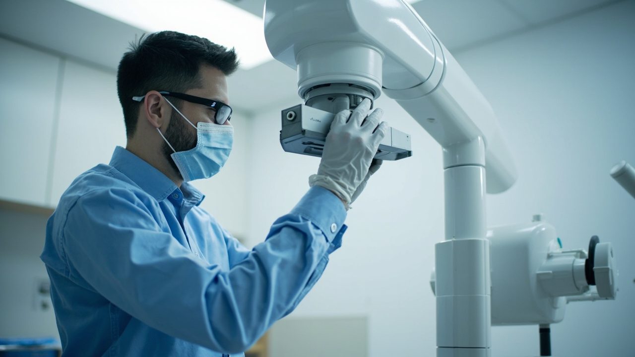
Monthly: Visual Inspection
- Inspect the tube head for oil leaks, unusual discoloration, or physical damage
- Check all cables and connections for wear or damage
- Verify that positioning arms move smoothly and lock securely
- Confirm that all indicator lights and displays function correctly
Quarterly: Quality Assurance Testing
Regular QA testing catches degradation before it affects clinical images:
- Output consistency testing: Use a dosimeter to verify that exposure output is consistent and within manufacturer specifications
- kVp accuracy: Verify with a kVp meter that actual output matches selected settings
- Timer accuracy: Confirm that exposure times match selected values
- Collimation check: Verify that the beam is properly collimated and aligned
- Image quality phantoms: Use a standardized phantom to objectively assess resolution, contrast, and uniformity over time
Keep a log of all QA results. Trending data is far more valuable than individual measurements — a gradual decline in output that’s still within spec tells you a tube is aging and helps you plan replacement proactively.
Annual: Professional Service
- Schedule annual service visits with a qualified x-ray equipment technician
- Full electrical safety testing including leakage radiation measurements
- Calibration verification with certified test equipment
- Oil condition assessment for tube housing integrity
- Regulatory compliance review — ensure your equipment meets current state and federal requirements
When to Replace vs. Repair
Tube replacement is expensive, but so is poor image quality and equipment downtime. Consider replacement when:
- Output has decreased more than 15–20% from baseline despite normal technique settings
- Image quality issues persist after all other variables (processing, sensor, positioning) have been ruled out
- Intermittent failures are increasing in frequency
- The tube has reached the manufacturer’s estimated exposure count or age limit
- Repair costs approach 50% or more of replacement cost
The Bottom Line
X-ray tube maintenance isn’t glamorous, but it directly impacts both your practice’s bottom line and your patients’ diagnostic care. A tube that’s properly warmed up daily, operated within its duty cycle, monitored with regular QA testing, and professionally serviced annually can last significantly longer than one that’s neglected.
The investment in preventive maintenance is minimal compared to emergency tube replacement — both in direct costs and in the disruption to patient care. Build these practices into your daily routine, and your equipment will reward you with years of reliable service.
Have questions about your specific x-ray equipment maintenance needs? Contact your equipment manufacturer’s service department or a qualified dental x-ray technician for guidance tailored to your setup.