Radiation Safety Protocols in Modern Dental Practices: Best Practices for 2025
Radiation safety remains a fundamental concern in dental radiography, requiring continuous attention to evolving protocols, regulatory standards, and best practices. As imaging technology advances and exposure guidelines are refined, dental professionals must maintain comprehensive safety programs that protect patients, staff, and themselves while ensuring optimal diagnostic quality. This guide examines current radiation safety protocols, implementation strategies, and emerging considerations for modern dental practices.
Understanding Radiation Exposure in Dental Imaging
Dental radiographic procedures expose patients and operators to ionizing radiation, necessitating careful balance between diagnostic benefit and radiation risk. Modern digital X-ray systems significantly reduce exposure compared to traditional film-based methods, but proper safety protocols remain essential regardless of the technology employed.
The ALARA principle (As Low As Reasonably Achievable) serves as the foundation for all radiation safety protocols. This philosophy requires minimizing radiation exposure through technical optimization, appropriate patient selection, and proper equipment utilization while maintaining diagnostic image quality necessary for clinical decision-making.
Current exposure levels for dental radiography are measured in microsieverts (μSv), with typical intraoral X-rays delivering approximately 5-10 μSv per exposure. To put this in perspective, these doses are comparable to natural background radiation exposure received during a few hours of normal daily activities.
Regulatory Framework and Standards
Radiation safety in dentistry operates under federal, state, and local regulatory oversight. The Nuclear Regulatory Commission (NRC), along with Agreement States, establishes fundamental radiation protection standards, while individual states maintain specific licensing requirements and inspection protocols for dental X-ray equipment.
The National Council on Radiation Protection and Measurements (NCRP) provides scientific recommendations that inform regulatory standards and clinical practice guidelines. NCRP Report No. 177 specifically addresses radiation safety in dentistry, offering comprehensive guidance on equipment specifications, facility design, and operational procedures.
Professional organizations, including the American Dental Association (ADA) and American Academy of Oral and Maxillofacial Radiology (AAOMR), publish practice guidelines and position papers that translate regulatory requirements into practical clinical protocols.
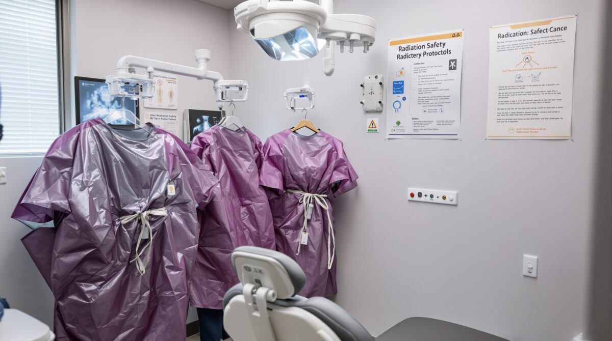
Essential Equipment Safety Features
X-Ray Machine Requirements
Modern dental X-ray equipment must incorporate multiple safety features to ensure compliance with current standards. Tube head design should include adequate filtration (minimum 2.5mm aluminum equivalent for machines operating above 70 kVp), proper beam collimation, and position-indicating devices (PIDs) that restrict beam size to the image receptor area.
Digital imaging systems offer inherent safety advantages through reduced exposure requirements, but proper calibration and quality assurance remain critical. Regular equipment testing ensures consistent performance and optimal radiation output characteristics.
Facility Design Considerations
Proper facility design minimizes radiation exposure to staff and adjacent areas. Primary barriers (walls, floors, ceilings) must provide adequate shielding based on workload calculations and occupancy factors. Secondary barriers protect against scattered and leakage radiation, with requirements varying based on distance and use factors.
Controlled areas, where radiation exposure may exceed certain thresholds, require restricted access and appropriate warning signage. Uncontrolled areas must maintain exposure levels below regulatory limits for members of the general public.
Personal Protection Equipment and Monitoring
Lead aprons and thyroid collars provide essential patient protection during radiographic procedures. Current recommendations specify lead equivalency requirements (typically 0.25-0.5mm lead equivalent) and proper coverage areas. Regular inspection and testing ensure continued protective effectiveness, with damaged protective garments requiring immediate replacement.
Operator protection relies primarily on distance and shielding rather than personal protective equipment. The six-foot rule establishes minimum distance requirements from the primary beam, while protective barriers or adequate distance provides secondary radiation protection.
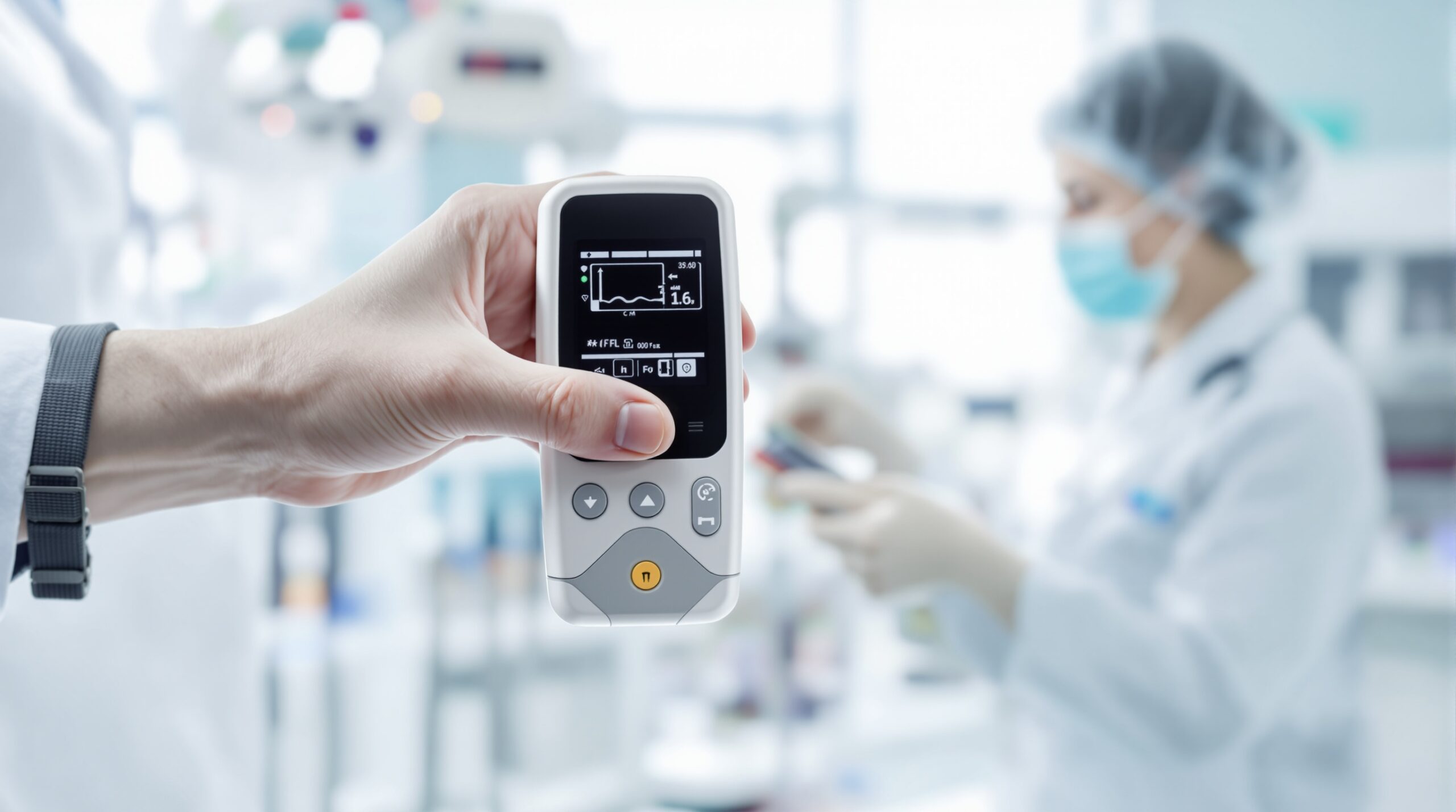
Dosimetry and Exposure Monitoring
Personal dosimetry monitoring tracks occupational radiation exposure for dental personnel who may receive significant exposure during routine duties. Modern dosimeters utilize optically stimulated luminescence (OSL) or thermoluminescent dosimetry (TLD) technology to provide accurate, sensitive measurements.
Monthly dosimetry exchange ensures timely exposure assessment and regulatory compliance. Dose records must be maintained for specified periods and made available for regulatory inspection and employee review upon request.
Area monitoring may be required in facilities with high-volume imaging or multiple X-ray units. Environmental dosimeters track facility exposure levels and verify shielding effectiveness over time.
Clinical Protocol Implementation
Patient Selection and Justification
Appropriate patient selection represents the first line of radiation safety. Clinical examinations should determine radiographic necessity based on patient history, symptoms, risk factors, and treatment planning requirements. Routine radiographic screening without clinical justification contradicts ALARA principles and may constitute inappropriate patient exposure.
Pregnancy considerations require special attention, with elective radiographic procedures generally deferred until after delivery. When radiographs are clinically necessary during pregnancy, standard protective measures, including lead aprons and thyroid protection, provide adequate fetal shielding for dental procedures.
Technique Optimization
Proper radiographic technique minimizes patient exposure while ensuring diagnostic quality. Optimal exposure parameters (kVp, mA, time) depend on patient size, dental structures being imaged, and image receptor characteristics. Higher kVp techniques generally reduce patient dose while maintaining image quality.
Image receptor selection impacts exposure requirements, with faster receptors (higher speed film or more sensitive digital sensors) enabling dose reduction. However, receptor selection must balance exposure reduction with diagnostic image quality requirements.
Collimation restricts the X-ray beam to the area of clinical interest, reducing patient exposure and improving image quality by minimizing scatter radiation. Rectangular collimation provides superior dose reduction compared to circular collimation, particularly for intraoral radiography.
Quality Assurance Programs
Comprehensive quality assurance (QA) programs ensure consistent equipment performance and radiation safety compliance. QA protocols include daily, weekly, monthly, and annual testing procedures that verify proper equipment function and safety feature effectiveness.
Equipment Testing Requirements
Daily quality assurance includes visual equipment inspection, darkroom cleanliness verification, and image quality assessment. Weekly testing may include image processor monitoring and digital sensor calibration checks.
Monthly QA procedures typically encompass more comprehensive equipment assessment, including exposure reproducibility, beam alignment verification, and safety feature testing. Annual inspections by qualified medical physicists or regulatory agencies ensure continued compliance with safety standards.
Documentation requirements mandate detailed records of all QA testing, equipment maintenance, and corrective actions. These records demonstrate regulatory compliance and provide evidence of proper safety program implementation.
Staff Training and Education
Effective radiation safety programs require comprehensive staff training covering basic radiation physics, biological effects, safety procedures, and regulatory requirements. Initial training should precede any involvement in radiographic procedures, with regular updates addressing procedural changes and regulatory modifications.
Competency Assessment
Regular competency evaluation ensures staff maintain appropriate knowledge and skills for safe radiographic practice. Assessment methods may include written examinations, practical demonstrations, and continuing education participation.
New employee orientation must address facility-specific procedures, emergency protocols, and safety responsibilities. Ongoing education maintains current knowledge of evolving regulations, techniques, and best practices.
Special Considerations for Pediatric Patients
Pediatric radiographic protocols require enhanced attention to radiation protection due to increased radiosensitivity and longer life expectancy. Exposure technique modifications, immobilization strategies, and parent/guardian involvement optimize image quality while minimizing radiation exposure.
Size-appropriate collimation becomes even more critical for pediatric patients, with beam limitation devices designed for smaller anatomical structures. Digital imaging offers particular advantages in pediatric dentistry through reduced exposure requirements and immediate image availability.
Emergency Procedures and Incident Management
Radiation safety programs must include emergency response procedures for equipment malfunctions, accidental exposures, and safety system failures. Clear protocols define immediate response actions, notification requirements, and investigation procedures.
Incident reporting ensures proper documentation of safety-related events and enables corrective action implementation. Regulatory notification requirements vary by jurisdiction and incident severity, but timely reporting demonstrates commitment to safety and regulatory compliance.
Emerging Technologies and Future Considerations
Advancing imaging technologies continue to influence radiation safety protocols. Cone beam computed tomography (CBCT) requires specialized safety considerations due to higher exposure levels and different radiation distribution patterns compared to conventional dental radiography.
Artificial intelligence integration in imaging systems may impact exposure optimization and quality assurance procedures. AI-driven exposure parameter selection and image enhancement could further reduce patient doses while maintaining diagnostic quality.
Regulatory evolution continues as new technologies emerge and scientific understanding advances. Staying current with regulatory changes and professional guidelines ensures continued compliance and optimal patient care.
Cost-Benefit Analysis of Safety Investments
Radiation safety investments extend beyond regulatory compliance to encompass patient confidence, professional liability reduction, and practice reputation enhancement. While safety equipment and training require financial commitment, the long-term benefits include reduced liability exposure, improved patient satisfaction, and enhanced professional credibility.
Modern digital imaging systems, despite higher initial costs, provide long-term benefits through reduced film and processing expenses, lower patient doses, and improved workflow efficiency. These factors often justify the investment through operational savings and enhanced practice capabilities.
Conclusion and Best Practice Recommendations
Effective radiation safety in dental practice requires comprehensive program implementation encompassing equipment maintenance, staff training, patient protection, and regulatory compliance. Success depends on leadership commitment, adequate resource allocation, and continuous improvement based on evolving standards and best practices.
Key recommendations include establishing written safety procedures, conducting regular training sessions, maintaining comprehensive documentation, and implementing robust quality assurance programs. Regular review and updating of safety protocols ensures continued effectiveness and regulatory compliance.
The ultimate goal of radiation safety programs extends beyond mere compliance to encompass optimal patient care through appropriate imaging utilization, minimal exposure levels, and maximum diagnostic benefit. By embracing these principles, dental practices provide exceptional care while protecting all individuals from unnecessary radiation exposure.