2024 ADA Radiation Safety Update: Lead Aprons and Thyroid Collars No Longer Recommended
The American Dental Association (ADA) made significant changes to radiation safety recommendations in February 2024, fundamentally altering how dental offices approach patient protection during X-ray procedures. The expert panel established by the ADA Council on Scientific Affairs determined that lead aprons and thyroid collars are no longer recommended for routine dental radiography.
The New ADA Position
After reviewing nearly 100 research articles, guidance documents, and regulations related to dental radiography, the ADA expert panel reached a clear conclusion: the use of lead abdominal aprons and thyroid collars during dental X-ray examinations provides no meaningful benefit to patient safety. This represents a major shift from decades of standard practice in dental offices across North America.
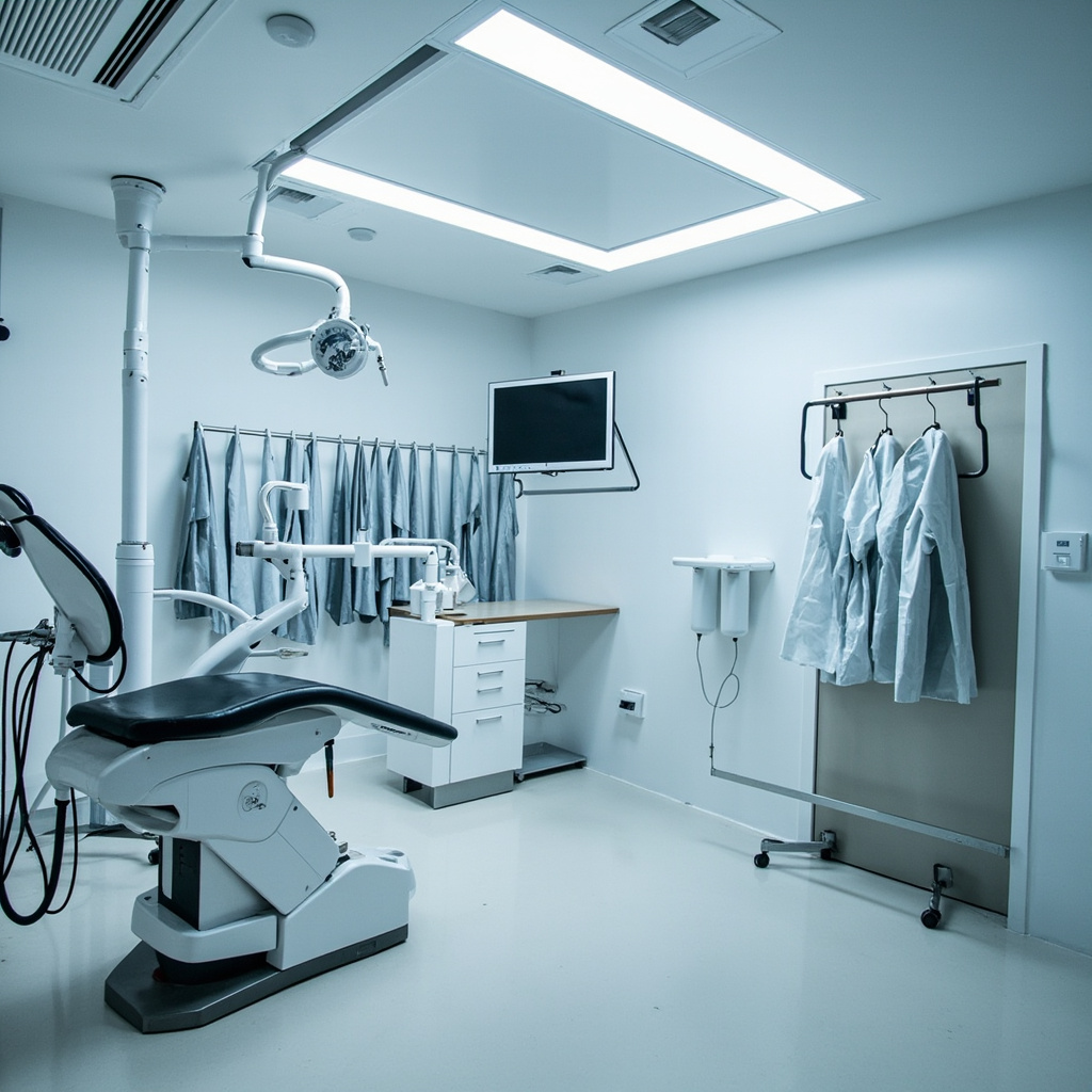
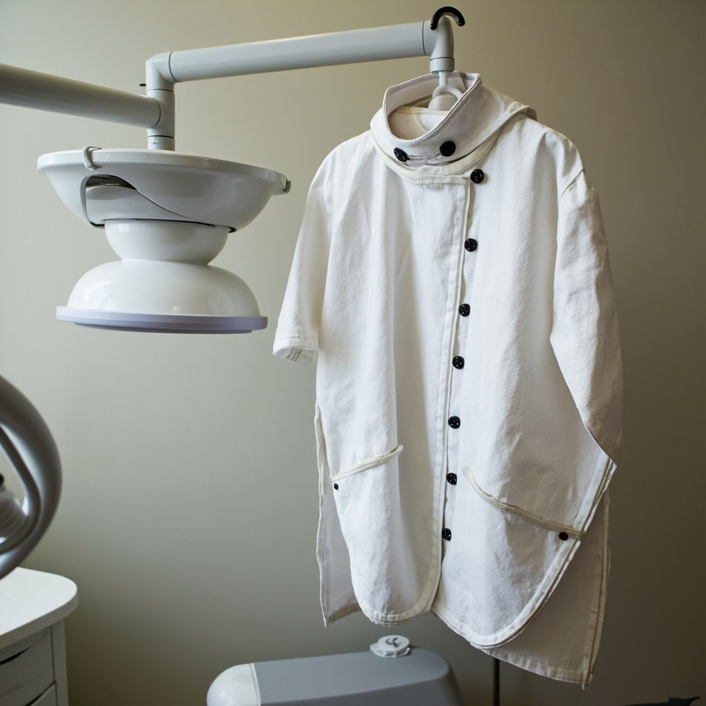
The recommendation applies to all forms of dental radiography, including intraoral bitewing and periapical X-rays, as well as extraoral panoramic and cone beam computed tomography (CBCT) examinations. The panel found that modern X-ray equipment and techniques have reduced radiation exposure to such low levels that additional shielding offers negligible protection while potentially compromising image quality.
Scientific Rationale Behind the Change
The decision was based on several key factors that have evolved since lead shielding became standard practice:
- Dramatically reduced radiation doses: Modern digital sensors and optimized exposure parameters have reduced patient radiation exposure by up to 90% compared to traditional film-based systems.
- Improved collimation: Contemporary X-ray units feature precise beam collimation that limits radiation to the area of diagnostic interest, minimizing scatter radiation.
- Digital image processing: Advanced image enhancement eliminates the need for retakes due to positioning errors or exposure issues.
- Evidence-based risk assessment: Current research shows the radiation doses from dental X-rays are so low that additional protection provides no measurable health benefit.
Dr. Raymond Cohlmia, Executive Director of the ADA, emphasized that “the radiation exposure from dental X-rays is incredibly small, and the additional protection from lead aprons and thyroid collars is essentially zero.” The panel’s analysis revealed that the doses involved are comparable to background radiation exposure from natural sources.
Implementation Challenges and State Variations
While the ADA has issued clear guidance, implementation varies significantly across jurisdictions. Some states maintain regulatory requirements that conflict with the new recommendations, creating compliance challenges for dental practices.
California’s Position: California law continues to require lead or lead-equivalent aprons covering reproductive organs for all patients undergoing dental X-ray examinations, including pregnant patients. The California Dental Association acknowledges the ADA’s updated stance but notes that state law supersedes professional recommendations until regulatory changes are implemented.
International Perspectives: The International Atomic Energy Agency (IAEA) maintains different guidance, still recommending thyroid collars “where the thyroid may be exposed to the main beam or to a considerable amount of scatter radiation.” This creates additional complexity for practices serving international patients or following global standards.
Practical Implications for Dental Practices
The transition away from lead shielding requires careful planning and staff education:
Staff Training Requirements
- Update radiation safety protocols and documentation
- Train team members on the scientific rationale for the change
- Develop patient communication strategies to address concerns
- Review and update office policy manuals
Patient Communication
Many patients expect to receive lead aprons during X-ray procedures, having been conditioned by decades of standard practice. Dental teams must be prepared to explain the scientific basis for the change and reassure patients about the safety of modern digital radiography. Clear communication about radiation dose comparisons can help patients understand that dental X-rays pose minimal risk.
Documentation and Liability
Practices should document their adherence to current ADA guidelines while ensuring compliance with applicable state regulations. This may require maintaining lead shielding for jurisdictions where it remains legally mandated while following ADA recommendations where permissible.
Quality Assurance and Alternative Safety Measures
With the removal of lead shielding, other radiation safety principles become even more critical:
- ALARA Principle: As Low As Reasonably Achievable exposure times remain the foundation of radiation safety
- Proper Collimation: Ensuring X-ray beams are precisely limited to diagnostic areas
- Regular Equipment Calibration: Maintaining optimal exposure parameters and image quality
- Digital Sensor Maintenance: Proper care and calibration of digital receptors
- Staff Training: Ongoing education on positioning techniques and exposure optimization
These measures provide far greater radiation protection benefits than lead shielding while ensuring optimal diagnostic image quality.
Looking Forward
The 2024 ADA radiation safety update reflects the evolution of dental technology and evidence-based practice. As digital radiography continues advancing and exposure doses decrease further, the focus shifts from physical barriers to optimized techniques and equipment maintenance.
Dental practices should stay informed about regulatory changes in their jurisdiction while maintaining compliance with the highest applicable safety standards. Regular review of radiation safety protocols ensures patient protection while embracing the benefits of modern dental imaging technology.
The transition away from lead shielding represents a significant milestone in dental radiology, demonstrating how scientific evidence can guide practice improvements that benefit both patient care and operational efficiency.