kVp and mAs Optimization in Dental X-Ray Systems: Essential Exposure Parameter Settings Guide

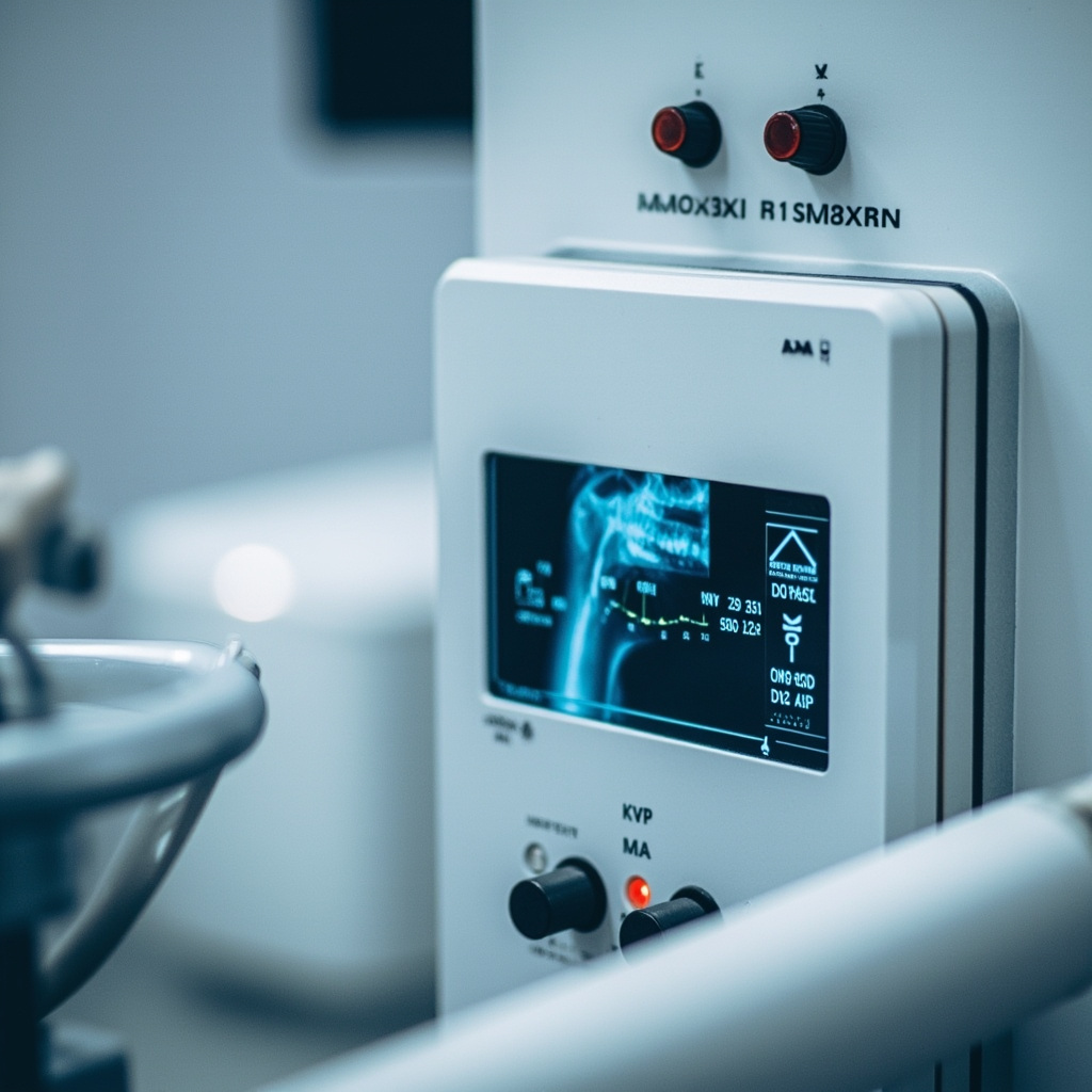
Proper exposure parameter optimization is critical for producing diagnostic-quality dental radiographs while minimizing patient radiation exposure. Understanding how to correctly adjust kilovolt peak (kVp) and milliampere-seconds (mAs) settings can significantly improve image quality and reduce retakes in dental practice.
Understanding kVp and mAs Fundamentals
Kilovolt peak (kVp) controls the penetrating power and energy of the X-ray beam, directly affecting image contrast and the ability to penetrate different tissue densities. Higher kVp settings produce X-rays with greater penetrating power, resulting in lower contrast but better penetration through dense structures like enamel and metallic restorations.
Milliampere-seconds (mAs) controls the quantity of X-rays produced, directly affecting image density or darkness. A 20% change in mAs is typically required to produce a visible change in image density on digital sensors.

Optimal kVp Settings for Different Clinical Scenarios
For general intraoral radiography, the optimal kVp range is between 60-70 kV. This range provides sufficient penetration for most dental structures while maintaining adequate contrast for diagnostic purposes. Patients with thicker bone density or those requiring imaging through metallic restorations may benefit from slightly higher kVp settings up to 80 kV.
When imaging pediatric patients, lower kVp settings of 60-65 kV are typically sufficient due to reduced tissue thickness. For bitewing radiographs in adults, 65-70 kVp provides optimal contrast between enamel, dentin, and carious lesions.
mAs Timing Considerations
Most modern dental X-ray units have preset mA values, making exposure time the primary variable for controlling mAs. Standard exposure times range from 0.1 to 1.6 seconds, depending on the imaging task and patient factors.
For anterior teeth, shorter exposure times (0.1-0.3 seconds) are typically adequate due to reduced tissue thickness. Posterior regions require longer exposures (0.4-0.8 seconds) to penetrate the increased bone density and tissue thickness.
The 15% Rule and Exposure Compensation
The 15% rule is a fundamental principle in radiographic exposure adjustment. When decreasing kVp by 15%, the mAs must be doubled to maintain equivalent image density. Conversely, when increasing kVp by 15%, the mAs should be halved.
This rule allows technicians to optimize image contrast while maintaining consistent exposure levels. For example, if standard settings are 70 kVp at 0.4 seconds, reducing to 60 kVp would require increasing exposure time to 0.8 seconds to maintain image density.
Common Exposure Errors and Corrections
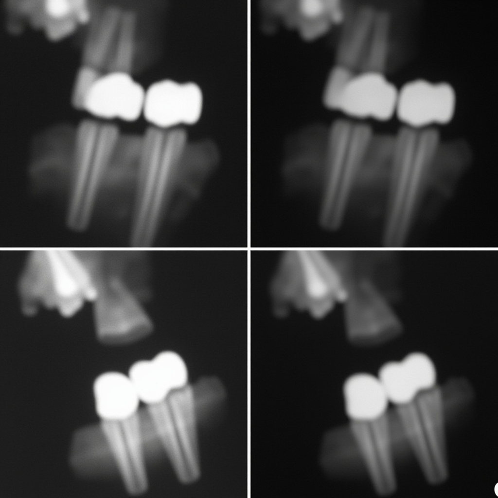
Underexposed images appear too light and lack sufficient density for diagnostic interpretation. This typically results from insufficient mAs or excessively high kVp settings. Increase exposure time or reduce kVp while compensating with increased mAs.
Overexposed images appear too dark and may obscure fine anatomical details. Reduce exposure time or increase kVp while reducing mAs accordingly. Digital sensors are more forgiving of overexposure than traditional film but optimal exposure still produces the best image quality.
Patient-Specific Adjustments
Large or muscular patients require increased exposure parameters, typically 25-50% more mAs than standard settings. Elderly patients with osteoporotic bone may require reduced exposure to prevent overexposure.
Patients with extensive metallic restorations may benefit from slightly increased kVp (75-80 kV) to improve penetration, with corresponding mAs adjustments to maintain proper density.
Quality Assurance Testing
Regular calibration of exposure parameters ensures consistent image quality and accurate exposure delivery. Annual testing should verify that actual kVp and mAs values match the control panel settings within acceptable tolerances.
Timer accuracy testing should confirm that exposure times correspond correctly to control panel selections. Digital imaging systems should be regularly calibrated to ensure optimal sensor response across the full range of exposure parameters.
Implementing standardized exposure protocols based on patient size, anatomical region, and clinical indication helps ensure consistent results and minimizes the need for retakes due to improper exposure selection.