The ADA’s 2026 Imaging Guidance Is a Hardware Problem: Service Factors That Keep Dose Low and Images Diagnostic

In January 2026 the American Dental Association published its first new dental X-ray recommendations in more than a decade, and the headline was blunt: dental radiographs are most effectively used in moderation, ordered only when clinically necessary, under the ALARA principle. Most of the coverage framed this as a prescribing decision for dentists. For those of us who service the equipment, there is a second, quieter message. Every exposure the practice does take should deliver the lowest reasonable dose while still producing a diagnostic image, and whether that actually happens is decided almost entirely by hardware you calibrate and maintain. Selection criteria decide whether to shoot. The machine decides how much dose that shot costs. This piece is about the second half.
ALARA Lives in the Beam, Not the Chart
A dentist can follow the selection criteria perfectly and still expose patients to far more radiation than necessary if the equipment is misconfigured. A round open-ended cone, a soft unfiltered beam, an over-driven exposure time, or a legacy-speed receptor can each multiply the dose of a clinically justified image several times over. The ADA guidance raises the stakes on this: if the profession is committing to fewer, more deliberate exposures, then each remaining exposure deserves hardware that is genuinely optimized. That optimization is a service deliverable, and it is measurable. When a technician walks into an operatory, the question is not “is this machine working?” but “is this machine producing a diagnostic image at the lowest dose it is capable of?” Those are very different inspections.
Rectangular Collimation: The Single Biggest Lever
If you change one thing on an intraoral unit to reduce dose, collimate the beam to a rectangle. A conventional round PID produces a circular field considerably larger than the rectangular receptor it is aimed at. All of that extra beam area irradiates tissue without contributing anything to the image, because it never reaches the sensor. Cropping the field to roughly the size and shape of the detector removes that wasted exposure. The reduction in irradiated tissue area is substantial, and because scatter is also reduced, image contrast can actually improve. It is one of the rare service changes that lowers dose and sharpens the image at the same time.
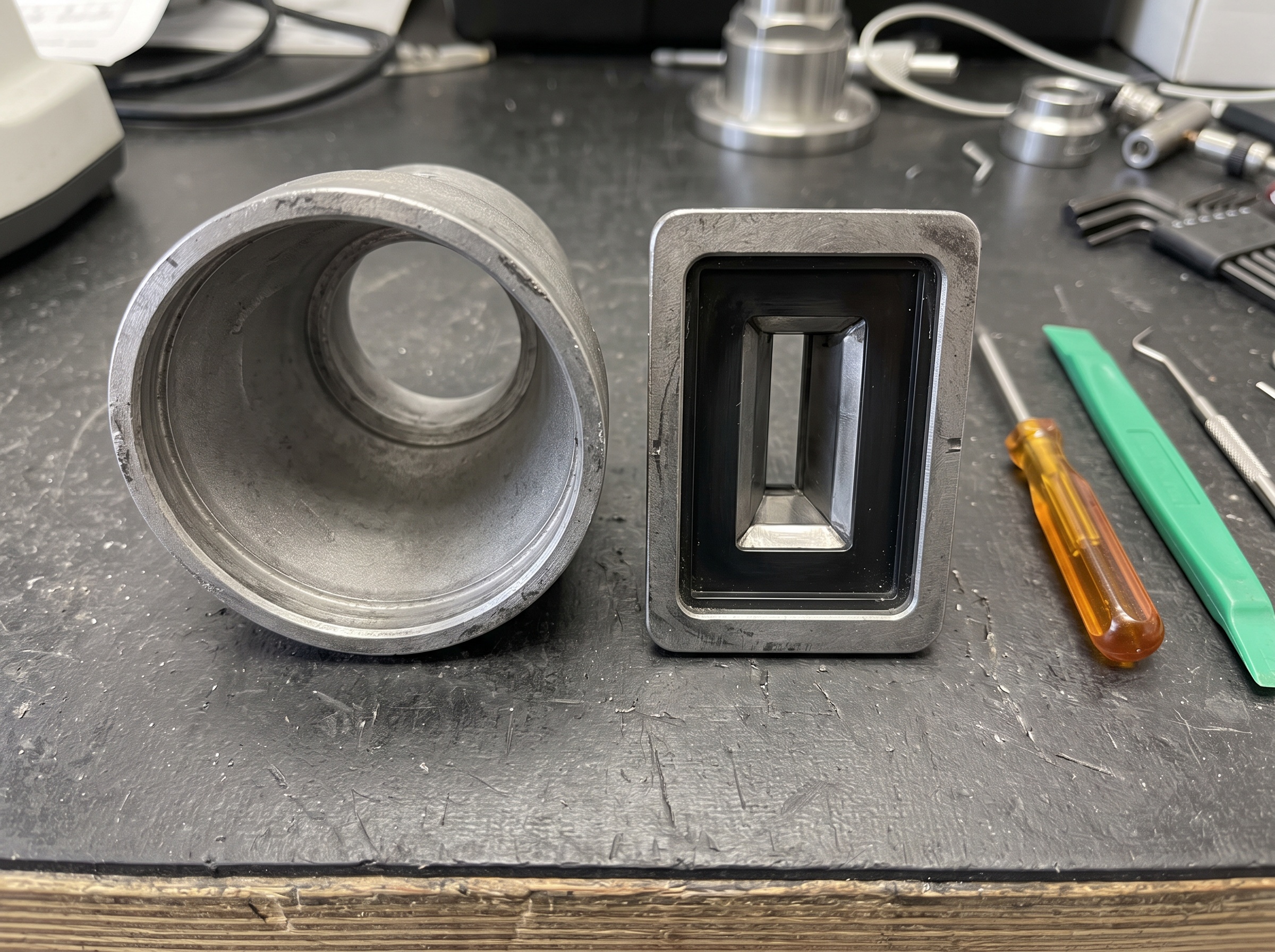
The catch is alignment. A rectangular field only works if it is squared to the receptor, so rectangular collimation and beam-alignment devices go together. As a technician, verify that a retrofit collimator is correctly indexed to the PID, that the practice is using a receptor-holder-and-aiming-ring system compatible with the rectangular field, and that staff understand why a tiny rotation of the tubehead now causes a cone-cut that a round beam would have forgiven. Pairing rectangular collimation with a proper alignment system is what turns the dose savings into diagnostic images rather than a rash of retakes.
PID Length, Alignment, and the Cone-Cut Tax
The position-indicating device is not just a spacer. Its length sets the source-to-skin distance, and a longer PID reduces beam divergence, which improves geometric sharpness and modestly lowers entrance skin dose for a given receptor exposure. During service, confirm the installed PID matches the unit’s design and the practice’s technique factors, that it is mechanically sound and not cracked or loose, and that any beam-alignment hardware seats squarely. Every cone-cut retake caused by poor alignment is a doubled dose for that image, so alignment integrity is a dose issue as much as an image-quality one. Verifying it belongs on every preventive-maintenance visit.
Detector Generation: Modern Sensors and PSP Versus Legacy Speed
The receptor sets the exposure floor. Modern digital solid-state sensors and current-generation photostimulable phosphor (PSP) plates require dramatically less radiation to form a diagnostic image than older film or the slow receptors many practices still run. If an operatory is exposing a fast modern sensor at technique factors inherited from a film era, it is over-exposing every image, which not only wastes dose but can saturate the detector and cost diagnostic quality. Part of the technician’s role is to confirm the exposure presets actually match the installed detector, and to flag when a practice running fatigued or outdated receptors would cut dose meaningfully by upgrading.
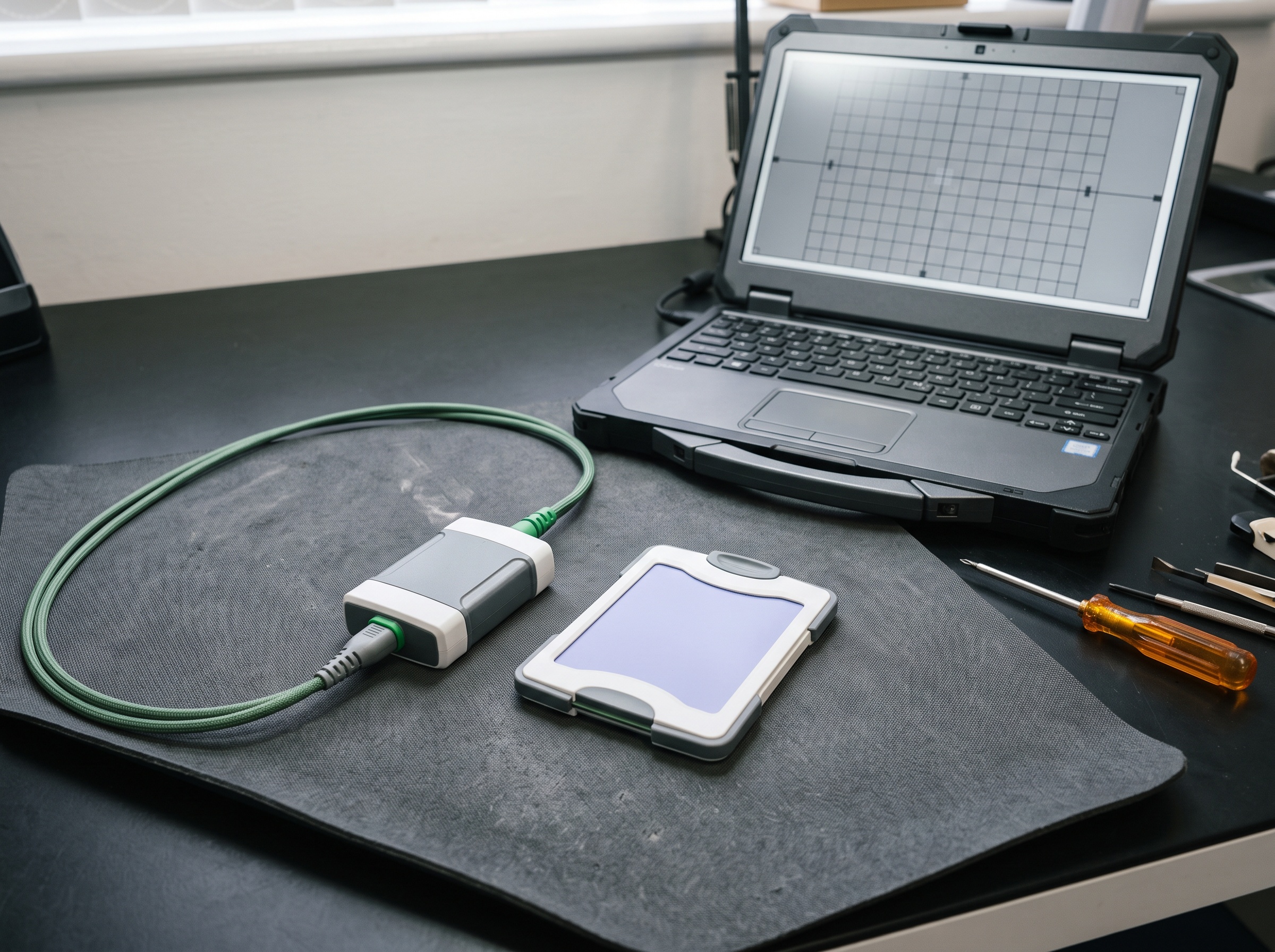
Detector condition matters as much as detector generation. PSP plates scratch, wear, and lose sensitivity with handling; a degraded plate tempts staff to raise exposure to compensate. Digital sensors carry dead or drifting pixels and need periodic flat-field and offset calibration so the software corrects them rather than the operator over-driving the beam. When you service the imaging chain, inspect the physical receptors, retire worn plates, and confirm the sensor’s calibration routine has been run to the manufacturer’s schedule. A well-maintained detector is what lets a low exposure still read as diagnostic.
kVp, mA, and Exposure Time: Calibration Is Dose Control
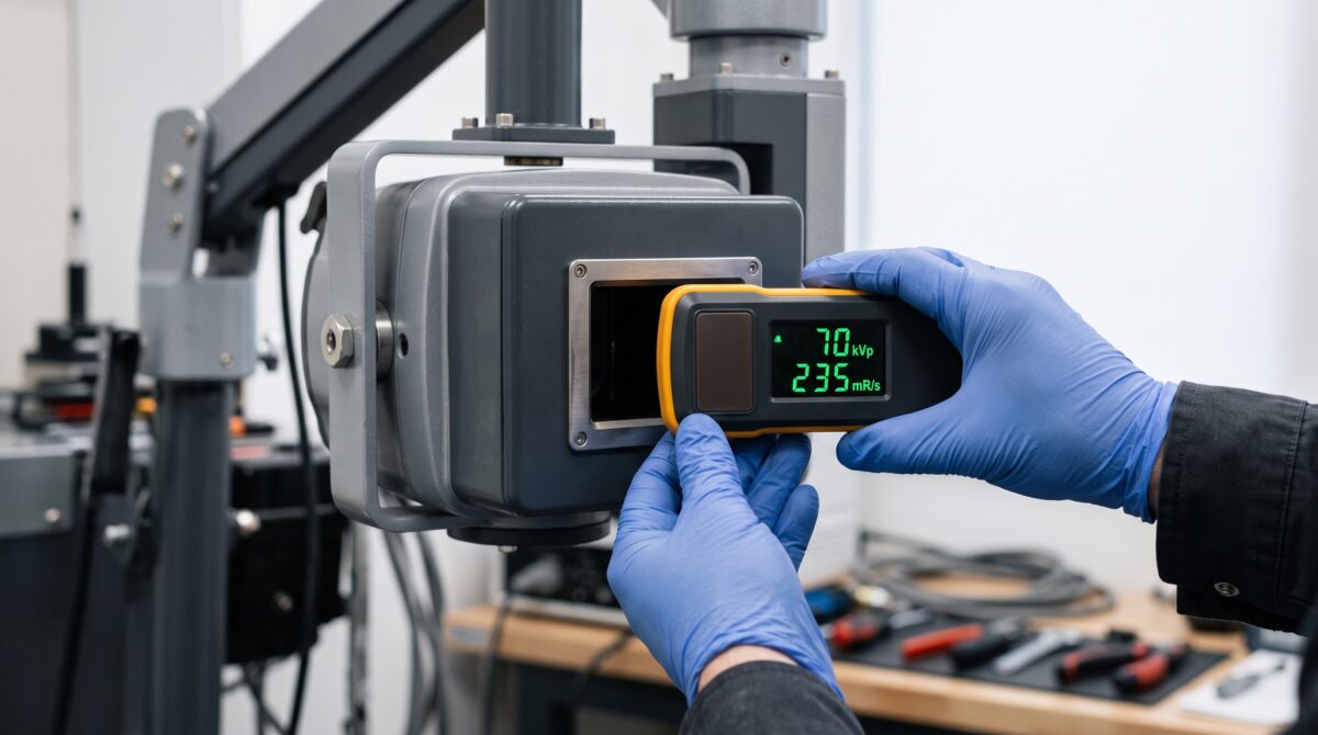
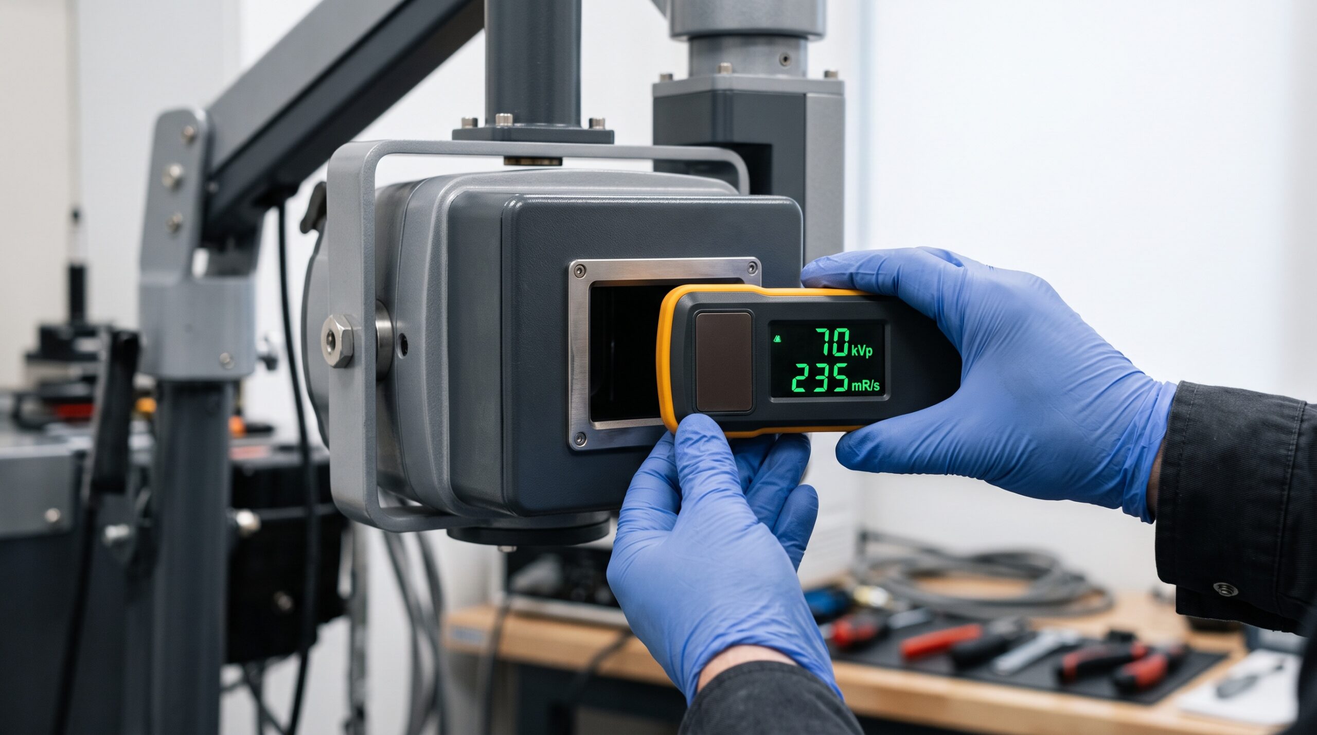
The exposure factors are where dose is set numerically, and they drift. A generator whose actual kilovoltage sits below the panel setting produces a softer, lower-energy beam that is absorbed more heavily by tissue and delivers more dose per useful photon. A timer that runs long, or tube current that reads higher than commanded, inflates every exposure directly. Because these errors are invisible at the operatory, they are exactly the kind of thing a technician must measure rather than assume. Using a calibrated dental X-ray meter, verify that measured kVp, mA, and exposure time track the panel settings within tolerance, and confirm the exposure is reproducible shot to shot at a fixed setting.
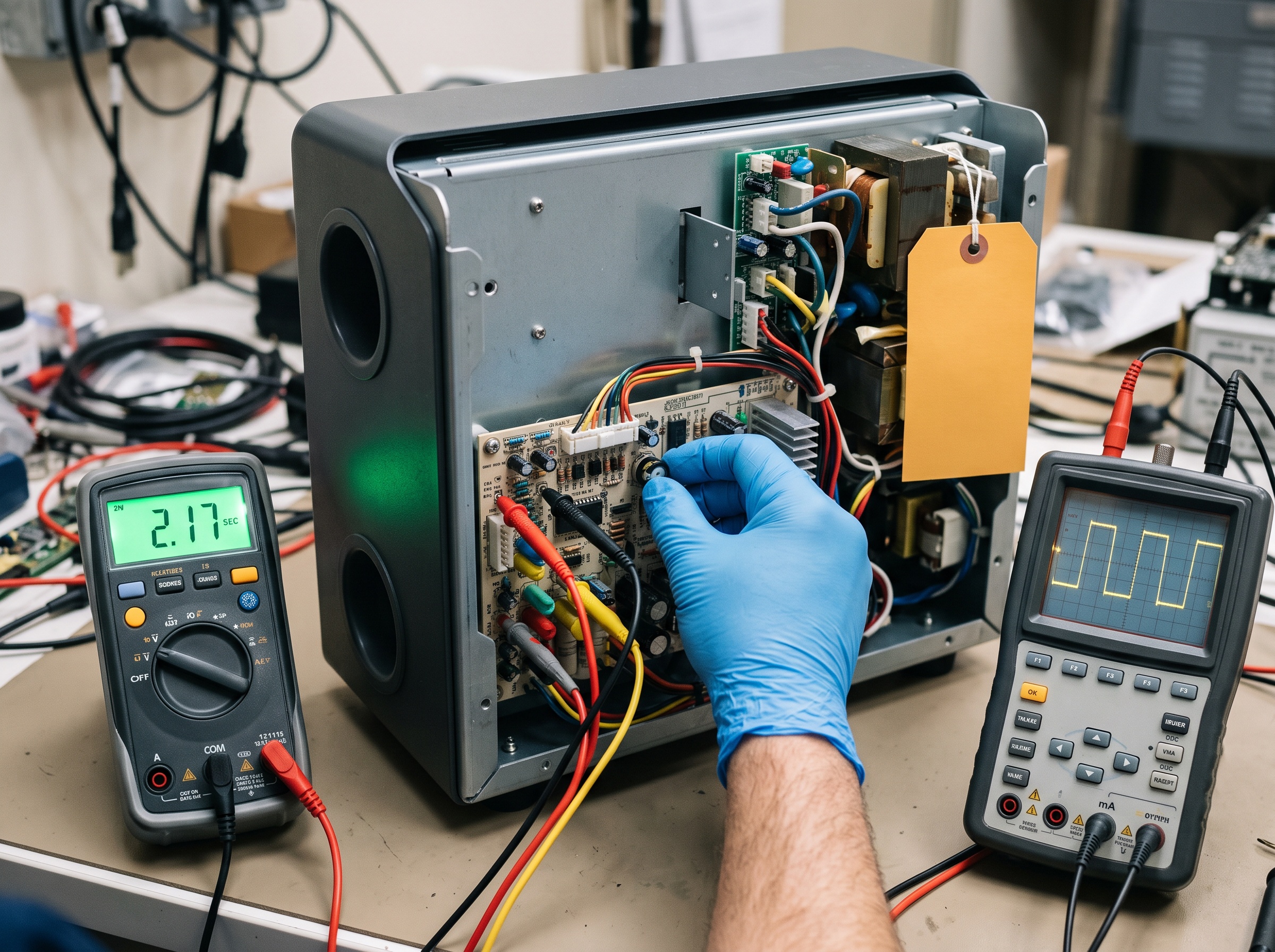
Reproducibility is its own dose safeguard. If identical settings produce swinging outputs, operators respond by defaulting to higher factors so the “weak” exposures still read, which raises dose across the board. Bringing timer, kilovoltage, and tube current back within specification lets the practice run the lowest factors that reliably yield a diagnostic image, which is the operational definition of ALARA at the panel.
Filtration, Half-Value Layer, and Tube-Output Consistency
Beam quality is a serviceable quantity, and it is easy to overlook because you cannot see it. Added aluminum filtration removes the low-energy photons that never penetrate to the receptor and instead deposit their entire dose in the patient’s skin. Regulations specify a minimum total filtration and a corresponding half-value layer (HVL), the thickness of aluminum that cuts beam intensity in half, as a proxy for adequate beam hardening. A tube with missing, cracked, or incorrect filtration produces a soft beam that spikes skin dose while doing little for the image. Measuring HVL with a meter and an aluminum step set, and confirming it meets the required minimum for the operating kilovoltage, is a core dose-reduction check that only a technician can perform.
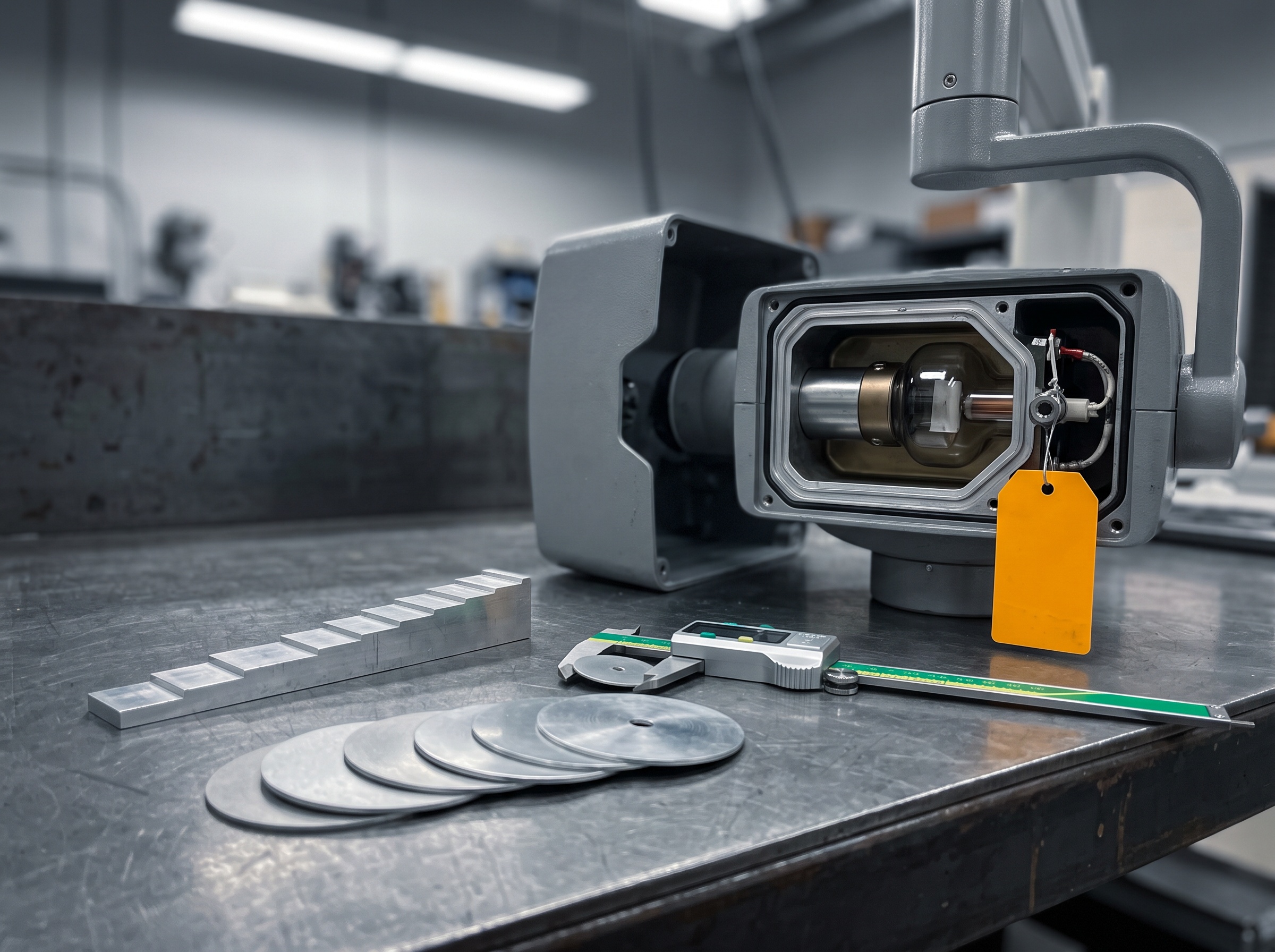
Tube-output consistency ties it together. As X-ray tubes age, output can drift and linearity between adjacent settings can degrade, so the same panel selection no longer delivers the same air kerma it did at install. Trending output over successive service visits catches a tube that is slowly forcing the practice into higher factors, and it flags a tube nearing end of life before it becomes an image-quality complaint. Consistent, correctly filtered, correctly calibrated output is the difference between a machine that merely functions and one that honors the ADA’s moderation-and-ALARA framing on every remaining exposure.
Monitoring, Prevention, and When to Call for Service
Make dose part of the maintenance rhythm, not an afterthought. On a scheduled preventive-maintenance visit, confirm rectangular collimation and beam alignment, inspect PID integrity, verify exposure presets against the installed detector generation, check receptor condition and detector calibration, and measure kVp, mA, exposure time, output reproducibility, and half-value layer against specification. Keep a log so you are trending these numbers over time rather than judging them in isolation; a slow drift is the earliest and cheapest thing to catch. Coach the practice that fewer exposures under the new ADA guidance only pays off in patient safety when each machine is tuned to deliver those exposures at minimum dose.
Some of this work requires instruments and expertise beyond a routine in-house check. If nobody has measured your kVp, timer accuracy, output reproducibility, or half-value layer with calibrated equipment, if you are retrofitting rectangular collimation and alignment hardware, if a tube’s output is trending downward, or if your jurisdiction requires periodic radiation-machine performance testing, that is the point to bring in professional service. The team at x-ray.support tests, calibrates, and optimizes dental X-ray equipment so that the profession’s move toward imaging in moderation is matched by hardware that keeps every justified exposure as low as reasonably achievable and still diagnostic.
Sources & further reading: