Common Digital Radiography Exposure Errors and Solutions
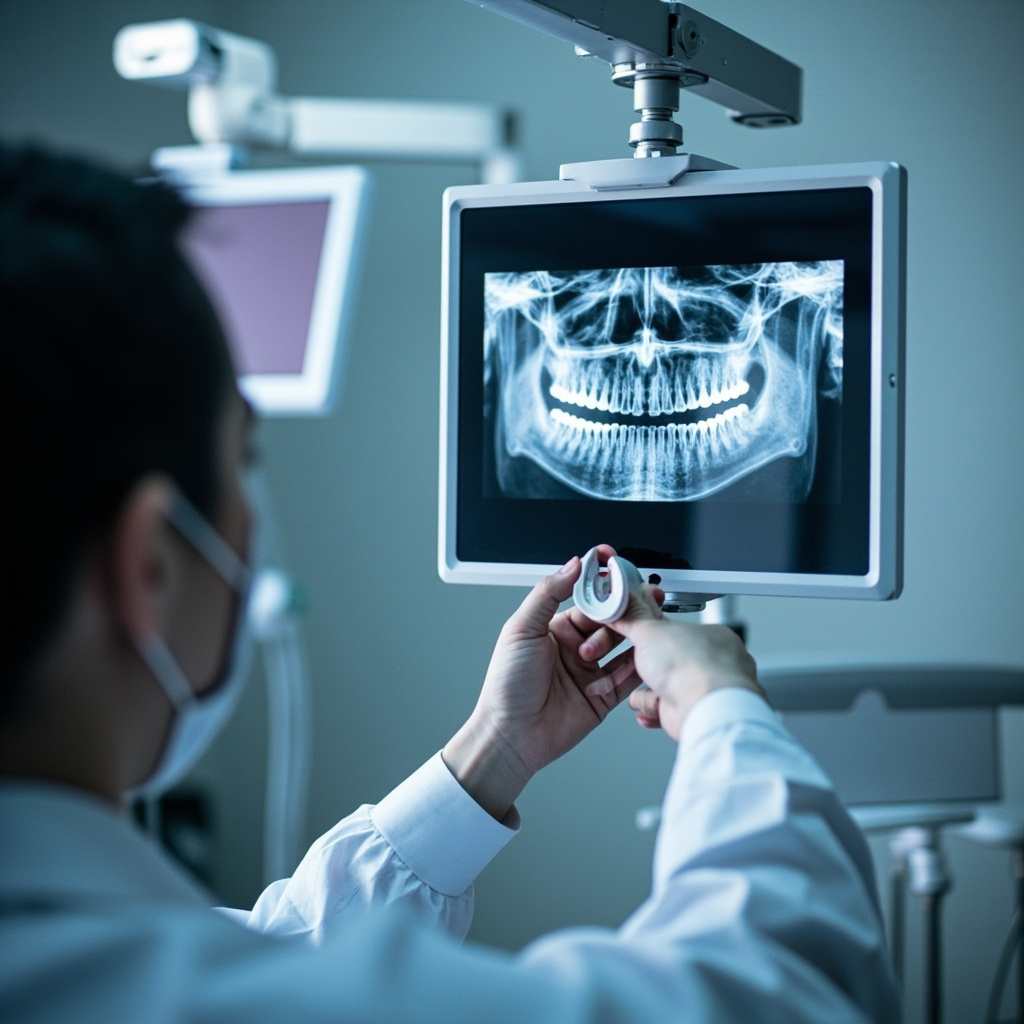
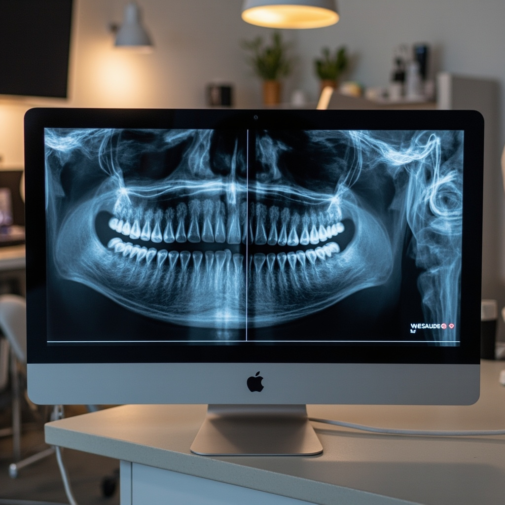
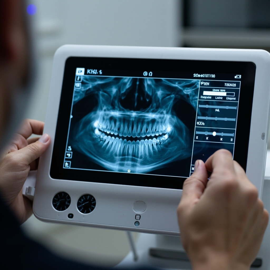
Digital radiography has revolutionized dental imaging, but exposure errors remain one of the most common technical challenges faced by dental practices. These errors can compromise diagnostic quality, necessitate retakes, and increase patient radiation exposure.
Understanding Exposure Parameters
Proper exposure in digital radiography depends on three critical factors: exposure time, kilovoltage peak (kVp), and milliamperage (mA). Unlike traditional film, digital sensors have different sensitivity characteristics that require careful adjustment of these parameters.
Most digital X-ray systems provide preset exposure values, but these defaults may not be optimal for every sensor type or clinical situation. Understanding how to manually adjust these settings is crucial for consistent image quality.
Common Exposure Errors
Underexposure
Underexposed images appear too light or dark (depending on the software display settings) and lack sufficient contrast for accurate diagnosis. This error typically results from:
- Insufficient exposure time
- Too low kVp settings
- Inadequate mA values
- Incorrect distance from the X-ray source
To correct underexposure, gradually increase exposure time in small increments (typically 0.1-0.2 seconds) rather than making large adjustments. For thicker anatomical areas like molars, consider increasing kVp rather than just extending exposure time.
Overexposure
Overexposed images show excessive density and poor contrast resolution. While digital sensors are more forgiving than film, overexposure can still degrade image quality and unnecessarily increase patient radiation dose.
Signs of overexposure include:
- Loss of anatomical detail in dense structures
- Reduced contrast between tissues
- Artifacts in the image processing
Systematic Troubleshooting Approach
When encountering exposure problems, follow this systematic approach:
Step 1: Verify Equipment Settings
Check that the X-ray machine settings match your sensor specifications. Many manufacturers provide recommended exposure charts for their sensors that serve as starting points.
Step 2: Assess Patient Factors
Patient size, age, and anatomical density significantly affect required exposure. Pediatric patients typically require 25-50% less exposure than adults, while larger patients may need increased settings.
Step 3: Evaluate Technique Factors
Ensure proper sensor placement, adequate patient positioning, and correct cone positioning. Poor technique can necessitate retakes regardless of exposure settings.
Sensor-Specific Considerations
Different digital sensor technologies (CCD, CMOS, photostimulable phosphor plates) have varying sensitivity characteristics. CCD sensors generally require less exposure than PSP plates, while newer CMOS sensors offer improved sensitivity with lower radiation doses.
Always consult your sensor manufacturer’s exposure recommendations as a baseline, then adjust based on clinical results and image quality assessment.
Quality Assurance Protocol
Establish a regular quality assurance program that includes:
- Monthly exposure consistency testing
- Sensor performance evaluation
- Image quality assessment using standardized phantoms
- Documentation of exposure adjustments and outcomes
Regular monitoring helps identify equipment drift, sensor degradation, or technique variations that could affect exposure accuracy.
Conclusion
Mastering digital radiography exposure requires understanding the interplay between equipment capabilities, sensor characteristics, and patient variables. By implementing systematic troubleshooting approaches and maintaining consistent quality assurance protocols, dental practices can minimize exposure errors and optimize both image quality and patient safety.
Remember that every exposure should be as low as reasonably achievable (ALARA principle) while maintaining diagnostic quality. When in doubt, consult with your equipment manufacturer’s technical support team for sensor-specific guidance.