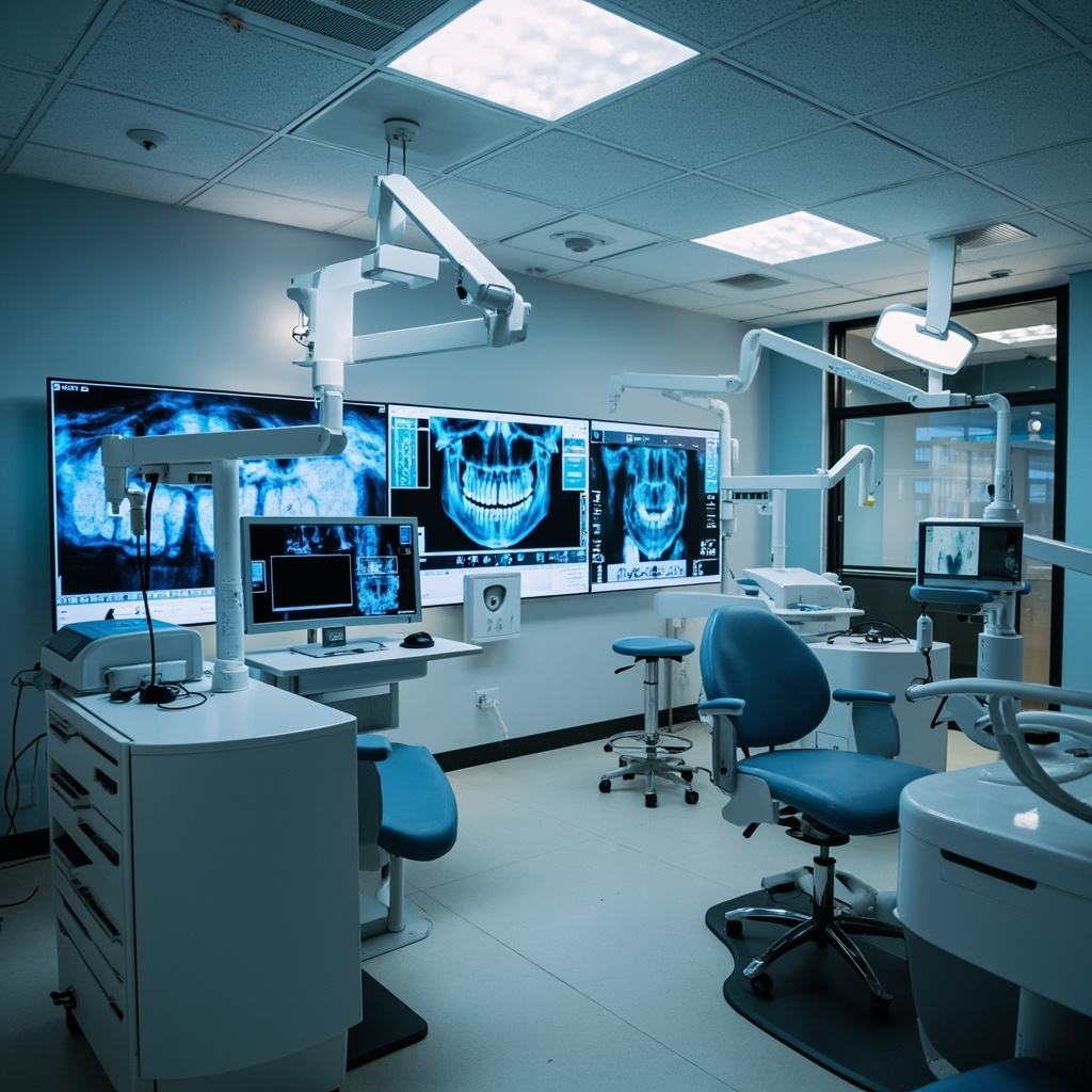
Dental imaging technology has evolved dramatically over the past two decades. While traditional two-dimensional X-ray remains the backbone of dental diagnostics, cone beam computed tomography (CBCT) has emerged as a powerful three-dimensional imaging modality that is transforming treatment planning across multiple specialties. Understanding when each technology is appropriate is essential for delivering optimal patient care while managing radiation exposure and costs.
What Is Traditional Dental X-ray?
Traditional dental X-ray encompasses several well-established imaging techniques that produce two-dimensional images. The most common types include periapical radiographs, bitewing radiographs, and panoramic radiographs (orthopantomograms). These modalities have been the standard of care for decades and remain indispensable for routine diagnostic tasks.
Periapical X-ray images capture the entire tooth from crown to root apex, making them ideal for evaluating individual teeth, detecting periapical pathology, and assessing root morphology. Bitewing radiographs excel at revealing interproximal caries and monitoring alveolar bone levels. Panoramic radiographs provide a broad overview of the entire dentition, jaws, temporomandibular joints, and surrounding structures in a single image.
What Is CBCT?
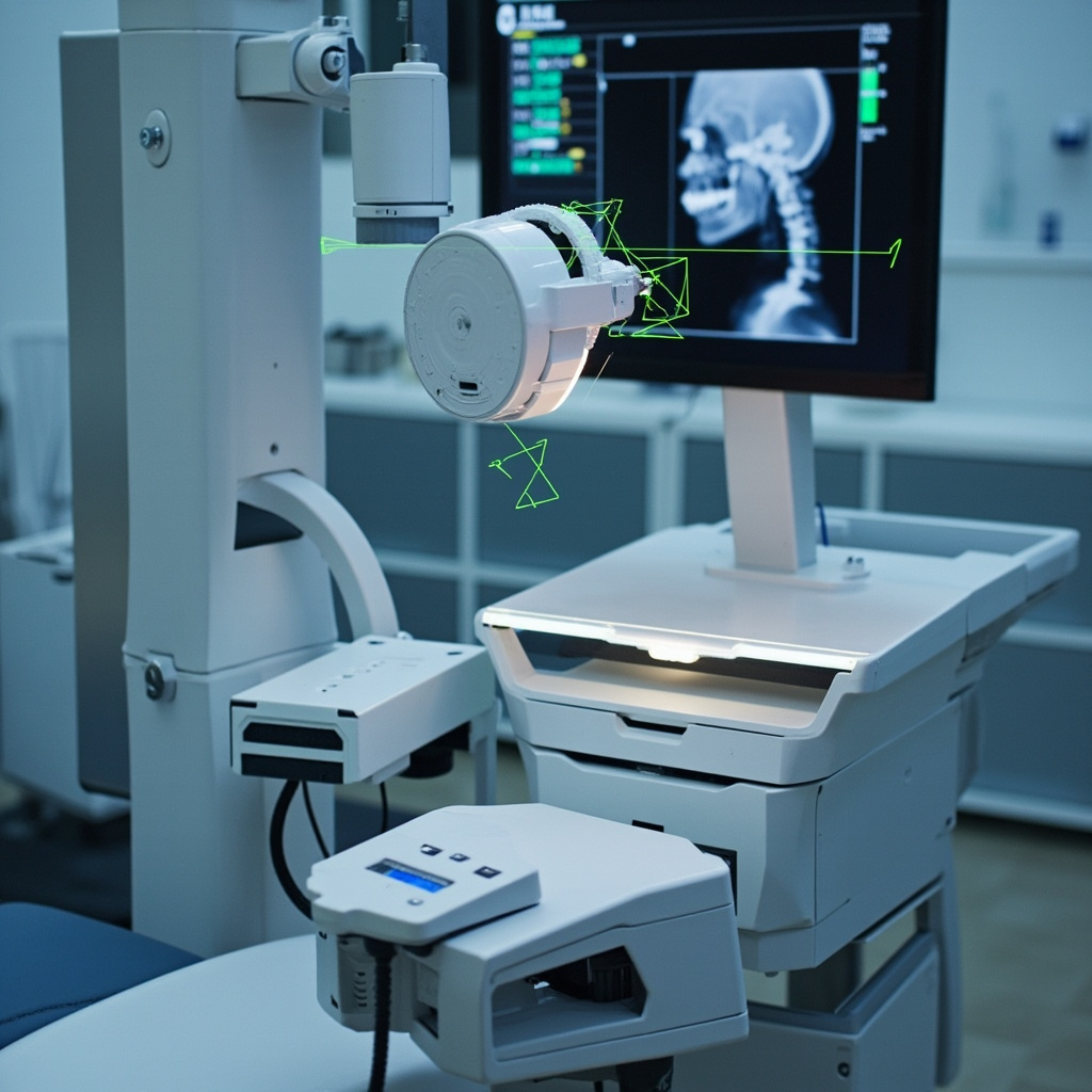
Cone beam computed tomography uses a cone-shaped X-ray beam that rotates around the patient’s head, capturing hundreds of projection images in a single scan. Sophisticated software reconstructs these projections into a three-dimensional volumetric dataset that clinicians can navigate in axial, sagittal, and coronal planes — plus generate cross-sectional slices at any angle.
CBCT scanners designed for dental use typically feature smaller field-of-view (FOV) options, faster scan times, and lower radiation doses compared to medical CT scanners. However, the radiation dose from even a small-FOV CBCT scan is significantly higher than that of a standard periapical or panoramic X-ray.
When Traditional X-ray Is the Right Choice
For the vast majority of routine dental examinations, traditional X-ray remains the appropriate first-line imaging modality. The following scenarios are well-served by conventional radiography:
- Caries detection: Bitewing radiographs remain the gold standard for detecting interproximal caries. Their high spatial resolution and low radiation dose make them ideal for periodic screening.
- Periodontal assessment: Bitewings and periapical X-ray images effectively demonstrate alveolar bone levels and can track periodontal disease progression over time.
- Routine endodontic evaluation: Periapical radiographs provide excellent detail for initial endodontic diagnosis, working length determination, and post-treatment follow-up in straightforward cases.
- General screening: Panoramic X-ray offers an efficient overview for new patient evaluations, orthodontic assessment, and third molar evaluation.
- Post-operative checks: Following routine restorative or surgical procedures, traditional X-ray is typically sufficient for monitoring healing.
Traditional X-ray benefits from lower cost per image, widespread availability, faster acquisition, and — most importantly — substantially lower radiation doses. The ALARA principle (As Low As Reasonably Achievable) demands that clinicians choose the lowest-dose imaging modality that can answer the clinical question.
When CBCT Is the Better Option
CBCT should be considered when two-dimensional imaging cannot provide the diagnostic information needed for safe and effective treatment. Key indications include:
- Implant planning: CBCT provides precise measurements of bone height, width, and density at the proposed implant site. It also reveals the exact location of critical anatomical structures like the inferior alveolar nerve canal, mental foramen, and maxillary sinus floor.
- Complex endodontics: Cases involving unusual root canal anatomy, suspected vertical root fractures, or resorptive lesions benefit enormously from three-dimensional visualization. CBCT can reveal additional canals that are invisible on periapical X-ray.
- Impacted teeth: When panoramic X-ray suggests a close relationship between an impacted third molar and the inferior alveolar nerve, CBCT clarifies the precise spatial relationship and guides surgical planning.
- Pathology evaluation: Large or complex jaw lesions, cysts, and tumors are better characterized with CBCT, which reveals their true three-dimensional extent and relationship to adjacent structures.
- Orthodontic and orthognathic surgery planning: Complex cases benefit from three-dimensional cephalometric analysis and airway assessment that only CBCT can provide.
- TMJ evaluation: CBCT offers superior visualization of bony components of the temporomandibular joint compared to panoramic X-ray.
- Trauma assessment: Suspected jaw fractures, root fractures, and dentoalveolar trauma may require CBCT when conventional images are inconclusive.
Radiation Dose Considerations
Radiation dose is perhaps the most important factor in choosing between these technologies. A single periapical X-ray delivers approximately 1–8 microsieverts (µSv), while a full-mouth series delivers about 35–170 µSv. A panoramic X-ray typically delivers 10–25 µSv.
By comparison, a small-FOV CBCT scan delivers approximately 20–100 µSv, while a large-FOV CBCT scan can deliver 70–600 µSv or more, depending on the unit and exposure settings. This means a single large-FOV CBCT can deliver radiation equivalent to several full-mouth X-ray series.
Every CBCT scan should be clinically justified — there must be a specific diagnostic question that cannot be answered by lower-dose imaging. Routine use of CBCT for screening purposes is not supported by current evidence-based guidelines from organizations such as the American Dental Association, the American Academy of Oral and Maxillofacial Radiology, or the European Commission.
Cost and Workflow Implications
Beyond radiation, practices must consider the financial and workflow implications. CBCT units represent a significant capital investment, typically ranging from $70,000 to over $200,000. Ongoing costs include software licenses, maintenance contracts, and staff training. Traditional X-ray equipment is considerably less expensive to acquire and maintain.
CBCT scans also require more time for interpretation. A single CBCT volume may contain hundreds of slices, and the clinician is responsible for reviewing the entire volume — including areas outside the region of interest. Incidental findings in CBCT scans are common and must be documented and managed appropriately.
Making the Right Decision
The decision between CBCT and traditional X-ray should always be guided by the clinical question at hand. Start with the lowest-dose imaging modality that can provide the necessary diagnostic information. If two-dimensional imaging leaves unanswered questions that are critical to treatment planning, then CBCT is justified.
Document your clinical rationale for ordering any imaging study, especially CBCT. Ensure that staff operating CBCT equipment are properly trained, that the unit is regularly calibrated and maintained, and that appropriate quality assurance protocols are in place. With thoughtful application of both technologies, dental practices can deliver the highest standard of diagnostic care while respecting the principles of radiation safety.