What Is the PID on a Dental X-Ray? The Position Indicating Device, Explained

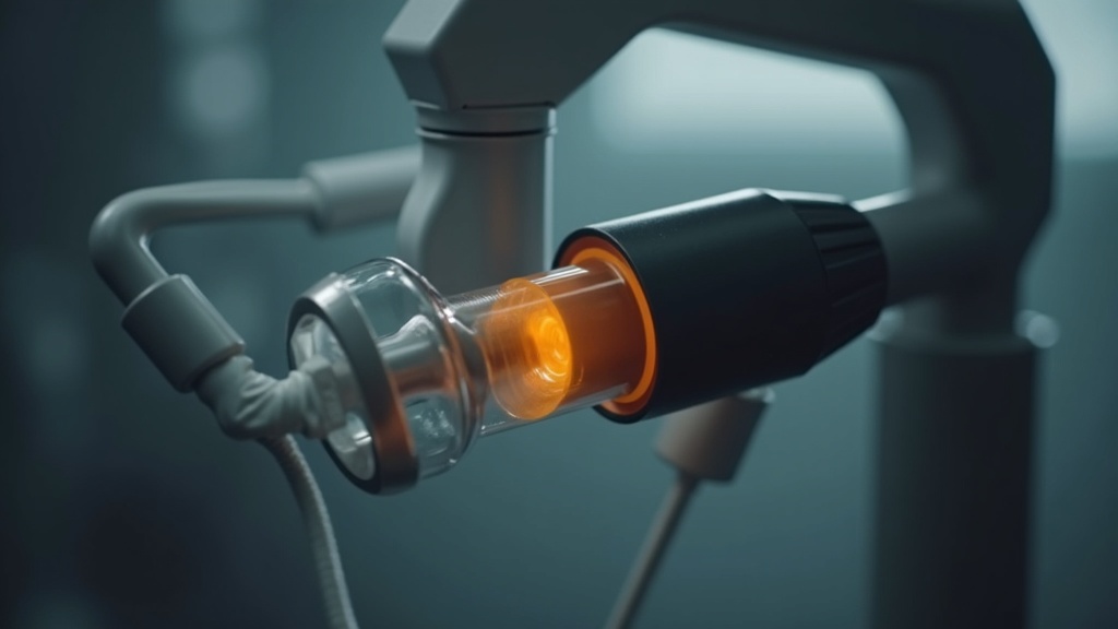
Ask three people in a dental office what the PID is and you may get three answers: “the cone,” “the pointy thing,” or a shrug. For a component that directly shapes both image quality and patient dose, it deserves a clearer explanation. PID stands for position indicating device — the open cylinder (or rectangle) that projects from the front of a dental X-ray tubehead. This guide covers what it actually does, the meaningful differences between types, and what a technician or equipment-minded practice should check.
PID Meaning: What the Position Indicating Device Does
The PID has two straightforward jobs, and one job it is often wrongly credited with.
Its first job is aiming. The PID indicates the position and direction of the X-ray beam so the operator can line the beam up with the receptor — the film or digital sensor — inside the patient’s mouth. That is where “position indicating” comes from.
Its second job is to establish the source-to-skin distance (SSD). Because the PID is a fixed length, it fixes the distance between the X-ray source and the patient’s skin, which in turn governs beam divergence and magnification.
The job people assume it does — limiting the size of the beam — is actually the collimator’s job. A lead collimator (a metal diaphragm with an aperture) sits inside the tubehead and restricts the beam. The PID then indicates where that already-shaped beam is going. On rectangular systems the two functions are combined, which is where much of the confusion comes from. Keep the distinction in mind: the collimator sizes the beam; the PID points it.
Round vs Rectangular PID: The Difference That Matters Most
If you take away one thing, make it this. A round PID produces a circular beam roughly 2.75 inches across at the patient’s face. An intraoral sensor is a small rectangle. That means a round beam irradiates a substantial area of tissue that never contributes to the image — it simply exposes the patient and generates scatter.
A rectangular PID (or a rectangular collimator retrofitted to a round one) trims the beam down to approximately the size and shape of the receptor. The dose reduction is significant — rectangular collimation can cut the irradiated area by more than half compared with a round field. This is why the American Dental Association and radiation-protection bodies specifically recommend rectangular collimation for intraoral radiography as a core dose-reduction measure. From a service standpoint, retrofitting a rectangular collimator or aiming ring to a round system is one of the highest-value, lowest-cost radiation-safety upgrades available to a practice.
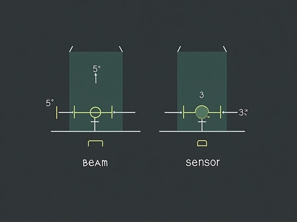
The trade-off is technique sensitivity: because a rectangular beam is only slightly larger than the sensor, alignment has to be good, or you get a cone cut (more on that below). The fix is not to abandon rectangular collimation — it is to use beam-alignment devices that lock the receptor, PID, and beam into register.
PID Length: Short vs Long, and What It Does to the Image
PIDs commonly come in two lengths, usually described as short (about 8 inches) and long (about 16 inches). Length changes the source-to-skin distance, and SSD changes the image:
- A longer PID (longer SSD) reduces magnification and improves sharpness. The beam reaching the receptor is more parallel, so the image geometry is truer to life — a real benefit for the paralleling technique.
- A longer PID requires more exposure. Following the inverse-square law, moving the source farther from the receptor reduces intensity at the film, so exposure factors must be set for the correct SSD.
- A short PID magnifies more and needs less exposure, but at some cost to geometric accuracy.
The practical service point: the PID length and the machine’s exposure settings are a matched pair. Swapping a PID for a different length without revisiting technique factors is a common cause of consistently over- or under-exposed images after a “simple” parts change.
A Word on Pointed Cones
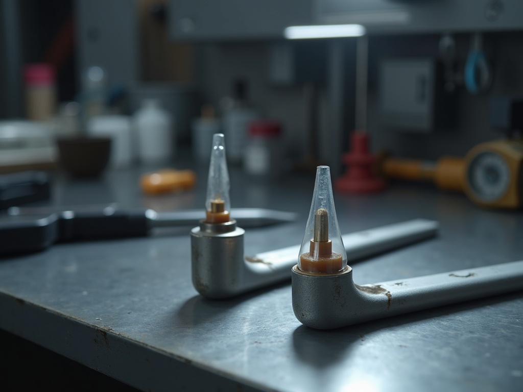
Older units sometimes still carry a pointed plastic cone. These should be retired. A pointed cone was once thought to help aiming, but the plastic actually generates significant scatter radiation as the beam passes through it, increasing dose to both patient and operator without any imaging benefit. Modern practice uses open-ended, lead-lined PIDs exclusively. Finding a pointed cone on a unit is an immediate flag to recommend replacement.
Cone Cuts and Other PID-Related Faults
The classic PID-related defect is the cone cut: a clear, unexposed curved (or straight, with rectangular systems) region on the radiograph where the beam missed part of the receptor. Diagnose it by cause:
- Misalignment — the beam was not centered on the sensor. With rectangular collimation this is easy to do by hand and is the reason beam-alignment holders exist.
- A PID not seated square — a loose or damaged mount lets the cone sit off-axis, so even a “correct” aim clips the field.
- Collimator/PID mismatch — an aftermarket rectangular collimator that is not properly indexed to the PID will throw the beam off consistently in the same direction, a useful diagnostic clue.
Beyond cone cuts, inspect the PID physically: cracks, a loose or wobbling attachment, a bent or dented cylinder, or any play in how it mounts to the tubehead. Because the PID sets both aim and SSD, a mechanically sloppy PID quietly degrades every image the unit takes.
What To Check, and When To Call for Service
For routine monitoring, work through a short list:
- Confirm the PID is open-ended and lead-lined, not a pointed plastic cone.
- Verify it is seated firmly and square, with no cracks or movement at the mount.
- Check that the collimation matches the receptor — ideally rectangular, with an alignment device in use.
- Confirm exposure factors correspond to the PID length in service.
- Watch the images: repeated cone cuts, unexplained density shifts after a parts change, or geometric distortion all point back toward the PID and collimation chain.
Aiming, alignment, and physical integrity a trained operator can monitor day to day. But anything involving the internal collimator, verification that the actual radiation field matches the light field, beam-alignment calibration, or a rectangular-collimation retrofit belongs to a qualified X-ray service technician with the right test tools. The PID looks like the simplest part on the machine — and precisely because it sits at the business end of the beam, getting it right is one of the most direct ways to protect image quality and keep patient dose as low as reasonably achievable.
Sources & further reading: